-
Life Sciences
.png)
Our solutions
-
Materials Analysis
.png)
Our solutions
Techniques
Applications
- Why Delmic?
-
Insights
.png)
Insights
-
Company
.png)
Company
Life Sciences
Accelerating the understanding of Type 1 Diabetes with FAST-EM

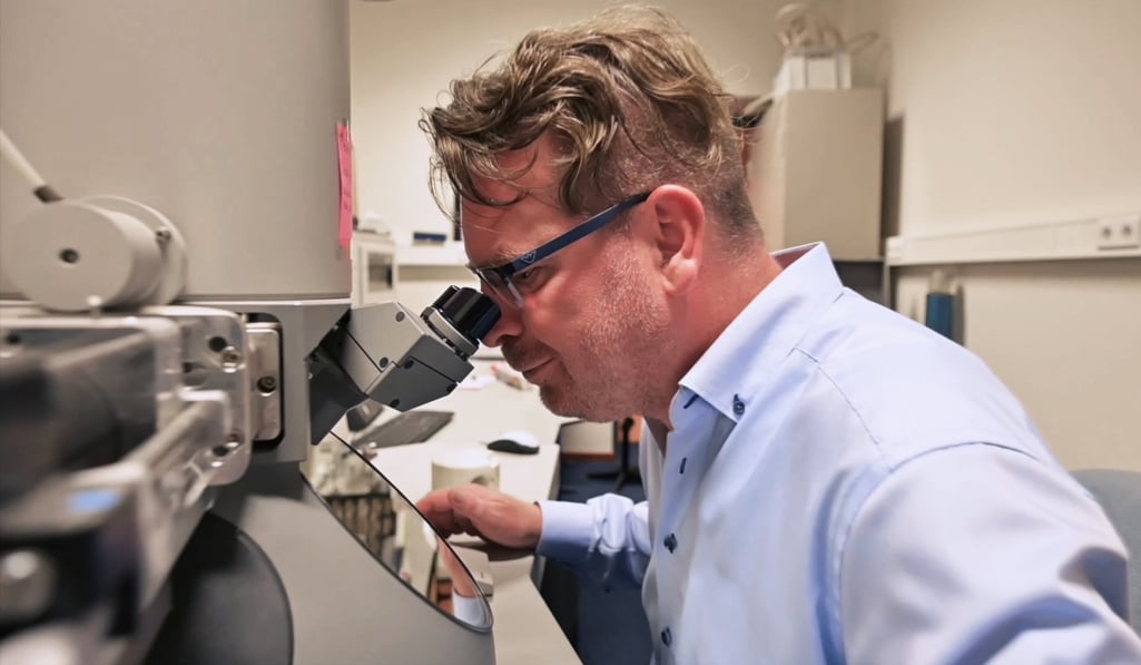
Dr. Ben Giepmans is a principal investigator of University Medical Center Groningen. His research is dedicated to Type 1 diabetes. Dr. Giepmans is intrigued by how biomolecules act together to control cell fate in health and disease. His team develops and implements new imaging techniques and probes for large-scale and multimodal microscopy. Particular focus is on Islets of Langerhans to help to understand trigger(s) and potential new therapies for Type 1 diabetes.
This is not commonly done in electron microscopy, it is a niche of our lab to complete a pipeline from a sample toward a large-scale image.
Dr. Giepmans points out two bottlenecks of traditional electron microscopy: the non-automated acquisition process and the low speed of the imaging. Dr. Giepmans mentions that the imaging process for an entire sample, usually conducted pixel by pixel, most of the time will yield a gigapixel image. Therefore, one image can take up from a few hours to over a weekend. Because of this, his lab currently mostly focuses on 2D images, and 2D analyses.
We typically don’t take 3D images because if you have a large area, it takes us 6 hours for one 2D image. If you want to have a thousand sections, it will take a year. If you now have a very fast microscope, we can go for 3D imaging within a week of such a section.
The powerful 64-beam scanning electron microscope (SEM), FAST-EM, is the perfect solution for Dr. Giepmans and his team since it can rapidly visualize large regions, at nanoscale resolution. Its ability to image much faster compared to a regular SEM and its high level of automation make FAST-EM a perfect system for imaging whole tissues or organs. One of the biggest benefits of the FAST-EM system, according to Dr. Giepmans, is its ability to speed up the acquisition process by imagining more pixels simultaneously.
Without the FAST-EM, we can not image 1 by 1-millimeter sample over the weekend, with the FAST-EM we will be able to do that over a couple of hours.
With FAST-EM it’s possible to acquire images in high resolution and create 3D datasets, which can provide insights and bring out a clearer picture of the Type 1 diabetes progression.
Acquired last year and currently installed at UMCG, the FAST-EM system is propitious in helping Dr. Giepmans and his lab with many projects, ranging from basic research to clinical use.


