-
Life Sciences
.png)
Our solutions
-
Materials Analysis
.png)
Our solutions
Techniques
Applications
- Why Delmic?
-
Insights
.png)
Insights
-
Company
.png)
Company
Life sciences
Advancing Scanning Electron Microscopy with FAST-EM

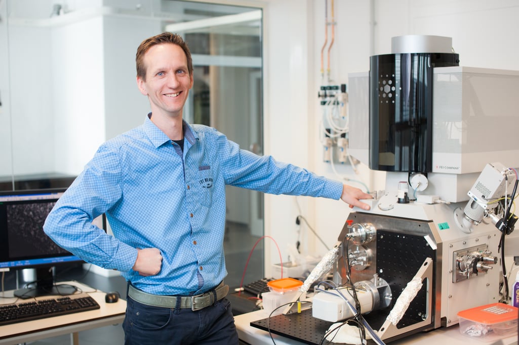
Dr. Jacob P. Hoogenboom is leading one of the ImPhys research groups at the Faculty of Applied Sciences at the TU Delft. The focus of his research group lies in the development of and research with new tools and techniques bridging the gap between light and electron microscopy.
The department of Imaging Physics integrates the fields of life sciences, healthcare and hightech industry. The group of Dr. Hoogenboom mainly focuses on developing innovative imaging technologies and instruments. These imaging systems are then put to use in groundbreaking research fields such as connectomics or cancer research. This dual function Dr. Hoogenboom facilitates creates collaboration and interaction between both the development and the application side of these techniques.
One of the most recently developed systems is the FAST-EM system. This multi-beam electron scanning microscope is developed at the TU Delft, in collaboration with Delmic, Technolution and Thermo Fisher. The main focus of this system is to overcome the throughput limitation. By operating with 64 beams scanning simultaneously, having a shorter dwell time and a fully automated workflow, the FAST-EM works 100 times faster than typical electron microscopes.
The detector that was developed for FAST-EM tackles that issue because it allows a faster imaging per pixel than regular electron detectors would do.
As an early adopter himself, Dr. Jacob Hoogenboom explored the different possible application fields for the FAST-EM system. In his opinion, the field of life sciences would benefit the most from a system such as FAST-EM. As stated above the system has the ability to lift the limitation of throughput. Some imaging facilities have a limitation in how many samples they can handle. FAST-EM would definitely benefit those facilities in processing more samples collected from faculties or hospitals.
One of the hardest research field in life sciences is large volume imaging of biological samples. With FAST-EM, the acquisition time is reduced from months to a few days, meaning that researchers have more time to image multiple specimens, multiple model systems, multiple animal brains, and at the end start comparative analysis earlier.
With FAST-EM, we can go to larger volumes. For instance, it could be used for a full model animal brain to map out all the neurons' connections.
Dr. Hoogenboom thinks the future of electron microscopy holds many new opportunities on both the technological as the biological side. The automation of the workflow will continue to take its course in both the imaging as well as the analysing part. In the end, data will probably be extracted from images automatically, speeding up many research pipelines. The world of imaging could go from imaging a single brain to imaging the whole central nervous system for example.
It offers new possibilities for my field of research - correlative light electron microscopy, especially in terms of neurons or system development. The electron microscope is now going to give volumes and areas that used to be the domain of light microscopy only.
As early adopters, as well as the initiators of the FAST-EM system, Dr. Hoogenboom's research team contributes to new opportunities for the research that is being done on the application side. The possibility to correlate these two modalities on larger scale length will bring in many research questions and requests for new technology, specifically for combining that data. The commercial multi-beam system at the TU Delft attracts users with biological life science-related questions and this, in turn, will bring the team into contact with their feedback and further requests.


