Dr. Sebastian Tacke works in the group of Prof. Dr. Stefan Raunser, Director at Max Planck Institute of Molecular Physiology (MPI), whose research focuses on the molecular understanding of fundamental cellular processes. To understand the mechanisms underlying these processes in healthy and diseased organisms in molecular detail, it’s essential to elucidate the structure and thus the function of the involved proteins and protein complexes.
Quite a few handling steps in the current cryo-EM sample preparation process can result in ice contamination of cryogenic samples. Dr. Sebastian Tacke and his colleagues faced this issue from the beginning of their research. During the first few cryo-focus ion beam (FIB)/scanning electron microscope (SEM) experiments, the researchers from the Raunser lab have already noticed that the throughput was very limited due to a low success rate of sample preparation. The cryogenic samples were more or less contaminated and the contamination was happening anytime throughout the whole cryo-electron microscopy (cryo-EM) workflow. To permanently solve this problem and further advance their research, they decided to establish a workflow, which circumvents the ice contamination, typically associated with the cryo-FIB sample preparation.
Ice contamination is of course particularly annoying when you have a limited amount of sample material.
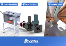
Tailored to ice contamination sources at different stages of sample preparation, Dr. Sebastian Tacke and colleagues started developing tools to protect their fragile samples from getting contaminated by crystalline and amorphous ice. The Cryo Workflow Tools, which are the prototype of Delmic’s CERES Ice Defence System, were created: the glove box (CERES Clean Station), the high vacuum cryo transfer (CERES Vitri-Lock) and the cryo shutter (CERES Ice Shield) . The glove box was designed to reduce the contamination (frost particles) during sample handling, the cryo-high vacuum transfer system aimed to provide maximum protection during all the transfers and the cryo shutter was focused on preventing parasitic ice growth from gaseous water molecules sublimation inside the cryo-FIB/SEM.
From my point of view, every cryo-FIB/SEM user who wants to automate the milling process will benefit from the cryo shutter (commercially as CERES Ice Shield). Only with it, we were able to run successful overnight sessions.
Since then the Raunser lab has been able to reduce the frost contamination by 50% and the contamination inside the cryo-FIB/SEM was not measurable anymore. The sample handling, pre-processing and milling processes in the cryo-ET workflow have improved dramatically with this patented innovation.
With these powerful tools, we were able to streamline the cryo-FIB/SEM automation process. Since there is no contamination visible, we were able to polish our lamellae automatically to a thickness of less than 150 nm on average, that was hitherto not possible with contamination.
Similarly to Dr. Sebastian Tacke, most of the researchers in biology struggle with ice contamination during their cryo-EM workflow, according to our research. Almost 40% of cryo samples become contaminated with ice and near a quarter of lamellae are rendered useless eventually due to ice contamination. Additionally, an astounding 43% of our respondents only discover this ice contamination when it’s too late and imaging at the cryo transmission electron microscope has already begun - a frustrating discovery because it often means that the sample can’t be used to acquire tomograms and the preparation work was all for nothing.
This is why we brought CERES Ice Defence System to the market: a unique solution, which we hope will help the researchers to stop wasting effort preparing precious samples only to have them destroyed by ice contamination. Instead, it will let them spend more time focusing on what really matters: increasing our understanding of the world around us with cryo-EM and getting that much closer to the next revolutionary breakthrough.
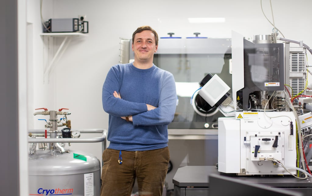
Photo: Dr. Sebastian Tacke in the Raunser lab. © Max Planck Institute of Molecular Physiology, Dortmund, Germany
This work is supported by the European SME2 grant № 879673
.svg "DELMIC")
.png)