Anna Bieber, Cristina Capitanio and Oda Helene Schioetz are PhD candidates in the groups of Prof. Dr. Wolfgang Baumeister, Prof. Dr. Brenda Schulman and Prof. Dr Juergen M. Plitzko respectively at the Max Planck Institute (MPI) of Biochemistry in Martinsried, Germany. They and their colleagues study all kinds of cellular processes with in situ cryo-electron tomography (cryo-ET). In the process of planning, designing and performing experiments they are constantly thinking about and trying out ways to improve the existing workflow to make correlative cryo-focused ion beam (cryo-FIB) milling and cryo-ET more robust, more effective and easier to use.
Research story of Anna Bieber and Cristina Capitanio
Anna Bieber and Cristina Capitanio are currently focusing on autophagy, an important cellular waste disposal and recycling system. Using cryo-fluorescence light microscopy (cryo-FLM), cryo-FIB milling and cryo-ET, they are characterizing all the steps that a cell needs to take to engulf cargo into so-called autophagosomes for degradation.
A big issue when they started correlative cryo-FIB milling and cryo-ET was the difficulty to assess the success of targeting a specific cellular structure. They would only find out if targeting was successful retrospectively, when they visualized the structure in the TEM or in the final tomogram.
We really wished for a way to check our lamellae for the target signal without having to do additional transfer steps, which would again increase the risk of contamination.
Even if the process of correlation (2D or 3D) was carried out as precisely as possible, it could be obscured by uncontrolled lamella bending or whole sample movements during FIB-milling. To be certain the region of interest (ROI) was present in the lamellae, they would have had to check the lamellae once more in a cryo-fluorescence microscope after milling– an error-prone and time-consuming procedure.
Delmic’s integrated FLM, METEOR, allows monitoring the presence of the target signal during and after milling within the FIB chamber. By using METEOR, Anna Bieber and Cristina Capitanio were able to check for the presence and precise location of the target signal in lamellae without having to transfer the sample to a separate cryo-FLM or waste time on them in the cryo-TEM.
METEOR has the potential to drastically improve our sample yield, especially when dealing with bright and abundant fluorescence targets. In this case, integrated FLM imaging can be coupled with automated FIB-milling. Several lamellae can be milled blindly and then be checked for signal in one go, either after rough or fine milling.
All automatically milled lamellae with no signal can be discarded, while all the lucky ones can be directly transferred to TEM for tomogram acquisition. If the target signal was less abundant, Anna Bieber and Cristina Capitanio would previously perform a 3D correlation of the FLM with the FIB image to target a specific structure. The lamella yield per milling session was, however, not necessarily higher.
By using METEOR they could image the final lamellae, quickly verify the presence and determine the exact position of the target signal on the lamellae and find the target structures easily in the TEM. In this case, a 3D correlation and “mill and check” approach, in which fluorescence is frequently inspected and the milling strategy is constantly adapted to keep the signal in the lamella, is the best option.
Anyone who wants to perform correlative cryo-FIB milling can benefit from an integrated FLM system. People with a well-defined and abundant target structure can increase their yield by using METEOR in combination with automation. Additionally, METEOR can bring improvements when targeting very rare and even novel structures.
Research story of Oda Helene Schioetz
Oda Helene Schioetz’s research focuses on elucidating structural information on exo- and endocytosis of vesicles in pollen tubule tips, as well as, obtaining structural details on integrins in focal adhesion sites. She spends lots of time on sample preparation for cryo-ET, specifically optimizing high pressure frozen (HPF) methods, FIB milling, and lift-out methods.
Before using METEOR, the correlation of HPF samples in Oda Helene Schioetz’ projects was limited to 2D due to the lack of fiducials and featureless surfaces.
However, METEOR aids in the correct 2D localization precisely through its transformation, so Oda Helene Schioetz doesn’t need to rely on correlating images anymore. Additionally, METEOR provides a method to check if the ROI is still present in the milled lamella throughout the milling process, eliminating the problems caused by the inability to do 3D correlation for HPF samples. The likelihood of hitting the target is therefore increased and, in turn, the overall sample yield.
METEOR has been helpful in guiding the milling of my HPF samples which makes my life much easier. I believe it’s a very useful tool, especially for those dealing with HPF samples where correlation would otherwise be limited to 2D.
Answering scientific questions is never an easy and straightforward process. It requires patience and critical thinking to figure out proper methods to gain insights. At Delmic we are driven to make microscopy an easy and accessible technique by developing user-friendly solutions which help researchers to get insights faster.
Anna Bieber and Cristina Capitanio said:
When we started our PhD the idea of a FLM microscope inside of the FIB chamber seemed such a simple solution, yet it was so distant from reality. Thanks to METEOR, it is wonderful to be finally able to check for signals at any time in the lamellae without any transfer steps and feel more sure about the presence of our target structures in the lamellae when we take them to the TEM for tomogram acquisition.
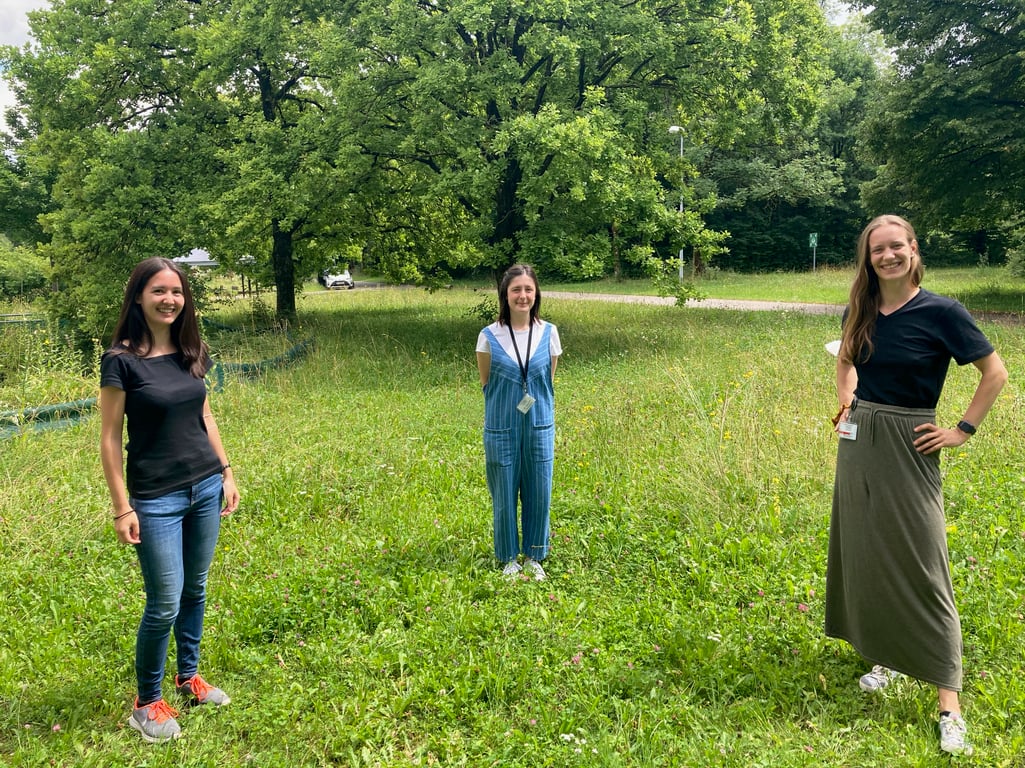
Photo: Anna Bieber (left), Cristina Capitanio (middle) and Oda Helene Schioetz (right)
This work is supported by the European SME2 grant № 879673
.svg "DELMIC")
.png)