Request pricing
micro and nanoplastics

Quantitatively characterize a variety of micro and nanoplastic materials with CL

Identify and characterise various plastics with fast, high resolution, easy-to-use cathodoluminescence solutions.

Read application note
What we can help you to achieve
  • Identify different plastics based on their characteristic spectra
  • Analyse particles ranging from nanometres into the microns or millimetres
  • Obtain correlative SEM and CL spectral maps 
  • Gain quantitative insights at high resolution
CL Plastics_Media w Description

Analyze intact plastic particles in high resolution

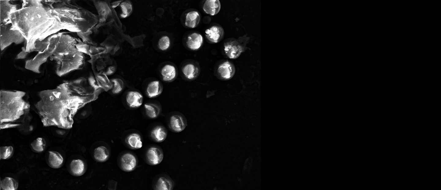
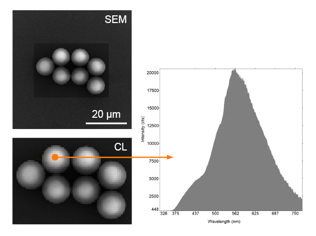
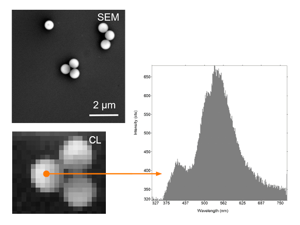
Plastics find wide use in our daily lives, with over 380 million tonnes of plastics being produced every year, of which about 40% is single use plastic1,2. Studies have also found that microplastics can act as vectors for heavy metal poisoning or adhere to chemical additives, which are then released and damage nearby tissue. Studying micro and nanoplastics has therefore become important as well as urgent. As an SEM-based high-resolution optical technique, cathodoluminescence (CL) imaging is promising since it can overcome many of the disadvantages in other popular techniques currently used for characterising plastic micro and nanoparticles, such as optical microscopy, Raman imaging, FTIR spectroscopy, gas chromatography, and SEM-EDX. CL is fast, easy to use, as well high resolution, capable of providing quantitative information and imaging particles of all sizes from nanometres into the microns or millimetres.

1. Lawnstarter, Plastic Statistics 2023      2. Plastics Europe, 2022

The general understanding of the mechanism of CL generation from plastics is bond cleavage by the electron beam, and the subsequent formation of luminescent polycyclic structures. The formation of radicals by the cleavage of aliphatic bonds may also lead to crosslinking, resulting in the generation of light under electron exposure. By performing CL spectroscopy, making sure to remain in the regime where the morphology of the particles is intact, we have performed proof of principle measurements to characterise a variety of micro and nanoplastic materials, for instance,  polystyrene, polyethylene, polypropylene, as well as a mixture from which polystyrene and Teflon could be identified.

What can you achieve with cathodoluminescence?

Our recent studies have identified CL imaging as a very promising fast, high-resolution technique for the characterisation of micro and nanoplastics. As shown in the image slider on the right side, the complete spectral information of 8μm polystyrene beads in the spectral range 300-800 nm was acquired for every pixel in the CL map, and overlaid with the SEM image. A peak is visible in this material at around 560 nm, with a shoulder at around 430 nm. Since the beads were large, they were imaged at 20 keV, which would excite the bulk. The high resolution of the technique is evident when imaging nanoparticles: we have found that the spectral map of 600 nm beads of the same material has approximately the same peak positions, but different peak widths and relative intensities. This is often the case for nanoparticles, whose properties can vary with size. Several plastics have been investigated and found to show characteristic CL peaks, demonstrating the promise of CL imaging for adding real value to the characterisation of micro and nanoplastics.

CL Plastics_MediaResize_5 keV CL Plastics_MediaResize_20 keV
Image slider showing a CL spectral map of polystyrene beads 8 μm in size and the spectral map of 600 nm beads of the same material

Use the right products to get the right results

Delmic CL solutions offer a range of powerful and user-friendly cathodoluminescence detectors, which can help you learn more about plastics. Choose the product fitting your research!

Talk to our expert

Want to learn more?

Talk to one of our experts to learn how our solutions can support you in your research.

Get in touch
Sangeetha avatar