Request pricing
Virology

Study viruses with integrated cryo-CLEM

Detect viruses and study their unique ultrastructure simultaneously by combining the power of light and electron microscopy in an unprecedentedly efficient cryo-ET workflow powered by our integrated cryo-correlative light and electron microscopy solutions.

Check our cryo-CLEM solutions
What we can help you to achieve
  • Identify and image viruses with in situ cryo-CLEM
  • Study replication mechanism of viruses at ease
  • Understand infected cells in a streamlined workflow
  • Efficiently prepare lamellas for further cryo-ET workflows
Header an integrated CLEM image showing Tupanvirus-infected amoeba cells

Understanding viruses in their near-native cellular environment

In the field of virology, cryogenic electron tomography (cryo-ET) is a novel technique which helps to study viruses in their near-native cellular environment and to reveal the interaction between the virus and its host, ranging from common viruses like influenza or rhinoviruses, to the current SARS-CoV-2. Many viruses evolve very rapidly due to the high mutation rate and short replication time, and cryo-ET can be particularly useful to examine the structural changes of the viruses during evolution.

However, the traditional cryo-ET workflow is more than complex with a significant number of transfer steps. The integrated cryo-correlated light and electron microscopy (cryo-CLEM) is the most advanced solution to date to simplify the traditional cryo-ET workflow, by allowing simultaneous coincident imaging with three beams: electrons, ions and photons. With effortless switching between fluorescent light microscope (FLM) and scanning electron microscope (SEM),  you can take full control of the in situ imaging and identify regions of interest (ROI) anytime during or after lamellae milling.

What can you achieve with integrated CLEM?

By integrating a FLM in your focused-ion-beam/ scanning electron microscope, we enable in situ cryo-CLEM and minimize transfer steps to establish a streamlined cryo-ET workflow. Not only are time and effort saved with the integration, but your precious viral samples are also less exposed to the risks of sample damage and ice contamination. With our CERES  Ice Defence System reinforcing the sample protection at every step of the workflow, your sample yield will be dramatically increased. 

  • Get viral lamellae in 7 simple fabrication steps
  • Acquire quantitative and three-dimensional data of viruses
  • Analyze replication mechanisms of viruses with high quality samples
Fluorescence image of a 100 nm thick ultra-thin section on ITO slide showing individual Tupanvirus particles High magnification SEM image of the same section, with no EM stain, showing individual Tupanviruses with the typical capsid and attached tail
(1) Fluorescence image of a 100 nm thick ultra-thin section on ITO slide using a 60X water immersion objective with an NA of 1.2, showing individual Tupanvirus particles both inside and outside the cell (2) High magnification SEM image of the same section, with no EM stain, showing individual Tupanviruses with the typical capsid and attached tail
MPI Martinsried_Website

METEOR has the potential to drastically improve our sample yield, especially when dealing with bright and abundant fluorescence targets. Anyone who wants to perform correlative cryo-FIB milling can benefit from an integrated FLM system...

Anna Bieber and Cristina Capitanio

-

Max Planck Institute of Biochemistry

Read the story

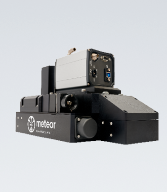
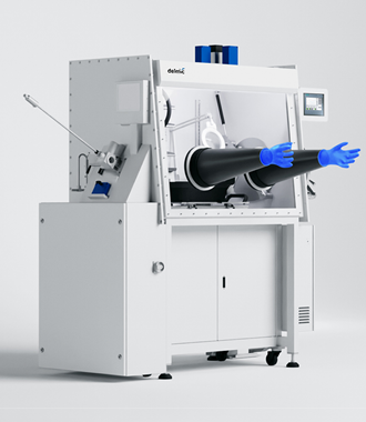
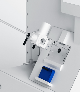
Choose the right product for your research

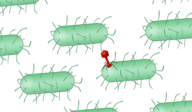
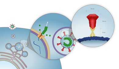
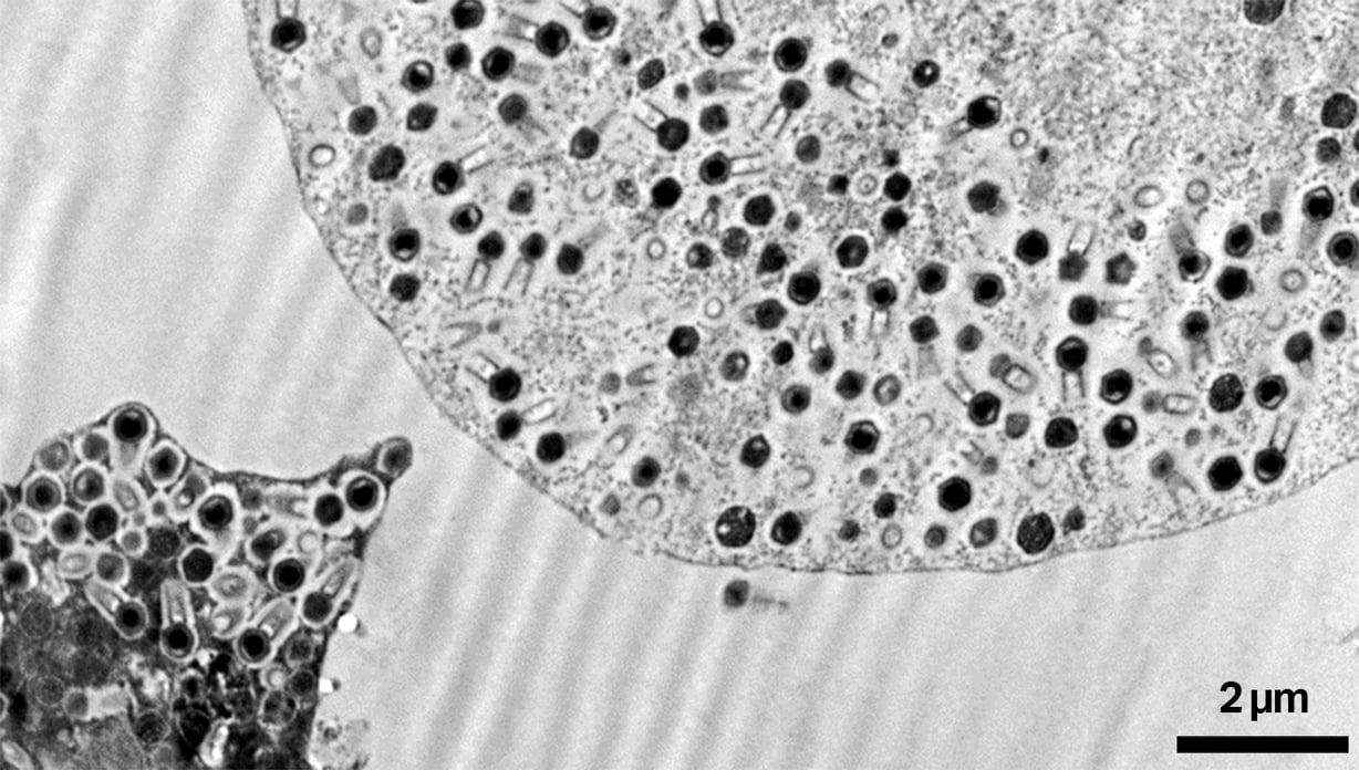
Delmic user-friendly and reliable cryo systems will help you to dramatically increase sample yield, obtain high quality, powerful data much faster and allow you to understand complex viruses better.

Talk to an expert

Want to learn more?

Talk to one of our experts to learn how our solutions can support you in your research.

Get in touch
CTA Bar - Marit - PNG