Choose the right product for your research
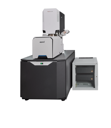
With out Fast Imaging solutions we aim to provide a stable, automated workflow based around minimal operator intervention.
Want to learn more about Fast EM?
Go to Fast Imaging technique page.svg "DELMIC")
.png)
Acquire more powerful insights to progress your research
Resolve optical properties at the nanoscale


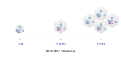
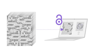
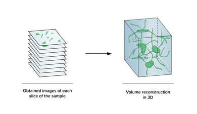
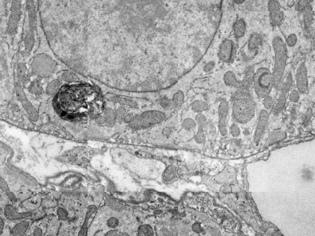
Unlock the potential of volume electron microscopy with higher throughput. Produce 3D data by imaging many subsequent sections of a sample with ease.

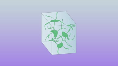
Three-dimensional electron microscopy or volume EM is a powerful method for understanding the architecture of tissues or organisms. This technique allows answering research questions in the fields of cell biology and neurobiology. Its high resolution is indispensable for visualization of the nanoscale details that define individual cells. Moreover, the ability to resolve the intricate structures of neurons at nanometer-range resolution is very useful for the field of connectomics which studies interactions between neurons in the brain.
To produce 3D data many subsequent sections of a sample need to be imaged and then reconstructed into a 3D representation for analysis. With increased throughput of faster electron microscopy, larger volumes can be imaged within a smaller amount of time. This opens the way to ever larger projects, such as imaging an entire hemisphere or even entire brains for connectomics. At the same time, high-throughput imaging enables comparative studies, which were previously too time-consuming to be feasible.
Most SEM techniques used for the collection of 3D datasets are challenging and require significant experience to reach their full potential. Unlike traditional SEMs, FAST-EM has a high level of automation which enables volume EM workflows with minimal operator supervision. Volumes of interest are rapidly collected, which enables neuronal tracing over large distances and a better understanding of cellular interactions in the brain and other tissues.
Dr. Jacob Hoogenboom
-Faculty of Applied Sciences, TU Delft