Request pricing
Cancer research

Get closer to understanding cancer with high resolution, large volume imaging

Understand disease progression faster and more efficiently, help to diagnose patients, and develop new treatments with the power of high-throughput electron microscopy. Create 3D models of whole organs and advance the fundamental understanding of cancer.

Learn about the solution
What we can help you achieve:
  • Get an overview of the entire ultracellular structure on the nanoscale
  • Image hundreds of sections with high level of automation and significantly faster
  • Collect nanoscale details while retaining the larger context of the sample, and understand how the tumor cell operates within the context of surrounding tissue
Cancer_FI_Media with Description

Study cancerous tissues in their entirety

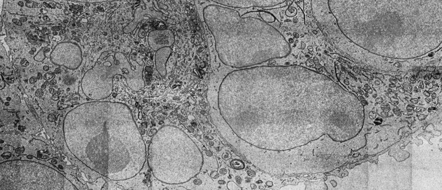
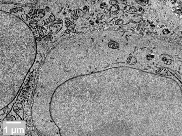
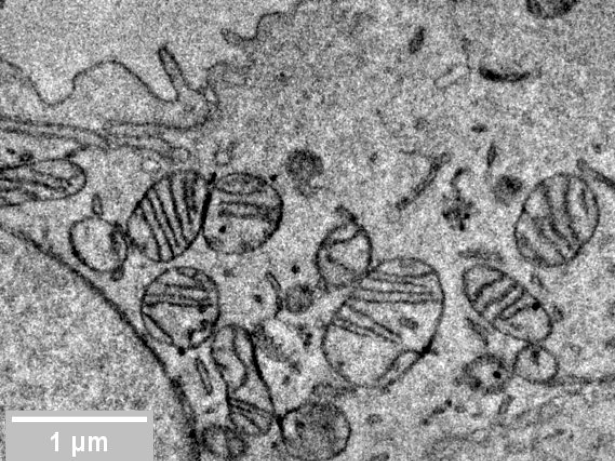
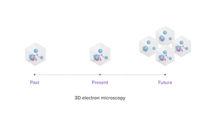
Cancer is a heterogenous disease which can acquire multiple hallmarks throughout its multistep development in human tumors. The six main hallmarks are evading growth suppressors, resisting cell death, sustaining proliferative signaling, enabling replicative mortality, activating invasion and metastasis, and inducing angiogenesis (sprouting of new blood vessels). When looking at both cellular organization and interactions between (cancer) cells and the tumor microenvironment, all these hallmarks can be observed by using high-resolution imaging. 3D electron microscopy is a powerful diagnostic technique in cancer research and one of the most accurate ways to get an overview of the entire ultracellular structure on the nanoscale.

With the use of 3DSEM, it is possible to identify different tumorigenesis hallmarks, enabling distinction between healthy tissue and tumor cells. However, traditional SEMs are often not able to handle the workload required for imaging thousands of sections necessary to create a 3D model. With FAST-EM this bottleneck is eliminated: this ultrafast multibeam SEM allows fast and automated acquisition of multiple sections in a matter of hours. As a result, it opens the way for comparative studies, where 3DEM volumes from different tumors or biopsies are compared to discover features common during tumorigenesis.

What can you achieve with FAST-EM?

Image large areas significantly faster and without the need for constant supervision. Acquire a large dataset to compare different samples or their parts, without compromising the image resolution. 

  • Acquire large datasets and compare samples of tissues from different patients, at different points in time, or biological material that was subjected to different treatments, without compromising image resolution
  • Obtain statistically relevant results with EM
  • Start answering fundamental research questions and make an impact in the scientific world
Cancer_FI_Media Resize_3 Cancer_FI_Media Resize_2
Breast cancer cell line derived from the mammary gland tissue of a mouse BALB/c strain.Sample fixed in 2% GA, stained using an enhanced osmium staining protocol (OTO-UL) and embedded in Durcapan resin.Sample courtesy of Radboud University Medical Center, Nijmegen.
Jacob Hoogenboom_IMG_9123-1

With FAST-EM, we can go to larger volumes. For instance, it could be used for a full model animal brain to map out all the neurons' connections

Dr. Jacob Hoogenboom

-

Faculty of Applied Sciences, TU Delft

Read customer story

Use the right products to get the right results

Delmic Fast Imaging solutions consist of FAST-EM, an ultra-fast automated multibeam electron microscope. Learn today what it can do for your research.

Talk to an expert
Fast Imaging

Want to learn more?

Talk to one of our experts to learn how our solutions can support you in your research.

Get in touch
Talk-to-Expert_Marit-de-Beer_Avatar