Request pricing
Cell biology

Identify and compare the smallest morphological differences in cells with high-throughput EM

Observe fine morphological details while retaining the large context of your sample. Acquire large-scale data and start comparing the sizes and shapes of organelles between cell types or different experimental conditions.

Learn about the solution
What we can help you to achieve
  • Use electron microscopy as a quantitative tool that provides answers to scientific questions rather than simply qualitative indication
  • Instead of gathering data only on the few regions of interest, capture large fields of view with EM
  • Collect large amounts of highly detailed data in a very short amount of time
Fig4bSR_RES

Get both complete overview and in-depth data at a macromolecular resolution

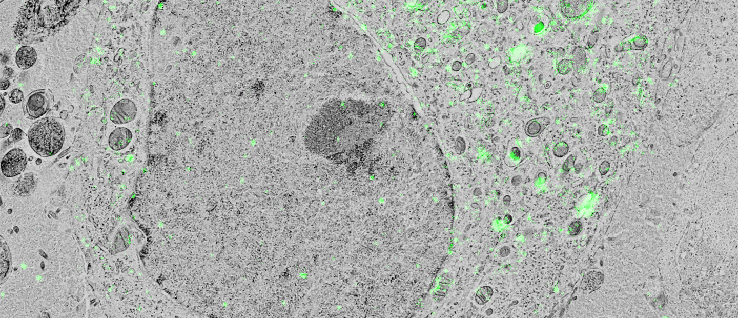
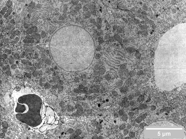
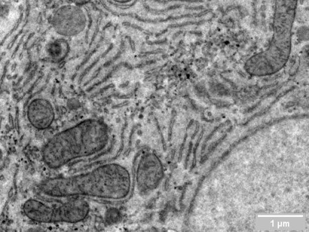
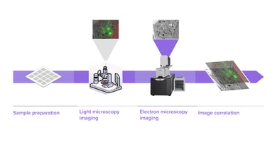
Researchers in cell biology rely on electron microscopy (EM) to answer their research questions since its high resolution is indispensable to visualize the nanoscale details that define individual cells. To analyze various aspects of the complex organization of cells, there is increasing demand to study the same sample at different length scales in biology. Correlative light and electron microscopy (CLEM) is a powerful technique that is used for this purpose. CLEM combines fluorescence microscopy (FM) with high-resolution electron microscopy (EM) to provide information about the composition and function of the sample. However, in traditional CLEM workflows, EM is often a limiting factor for the scope of data collected: with low throughput and limited field of view, careful selection of ROIs is needed to ensure the feasibility and success of a project. Moreover, the limited throughput of the workflow leads to the lack of context, which makes CLEM more of a qualitative technique rather than quantitative.

With high-throughput electron microscopy, CLEM workflows can be enormously simplified and can easily provide quantitative information to the researchers. Delmic’s ultra-fast multibeam scanning electron microscopy, FAST-EM, is capable of collecting megafield of the dimensions of roughly 250 µm x 250 µm, with 4 nm pixel size, within just 3 minutes. This means that you no longer have to compromise between the size and number of samples. Instead of gathering data only on the few labelled regions of interest, it is possible to capture large fields of view with EM and correlate it with the FM data. This type of quantitative analysis typically requires weeks or months to be completed with standard single-beam systems. With FAST-EM it could be completed within a few hours or days.

What can you achieve with FAST-EM?

FAST-EM is highly beneficial for large-scale electron microscopy, where high-resolution imaging is performed on large samples ranging from tissues or organs. Large-scale imaging provides both nanoscale information to analyze subcellular types and the context needed to understand the distribution of cell types within tissues or organs. With FAST-EM you will be able to:

  • Autonomously acquire large datasets 
  • Perform comparative studies of sizes and shapes of organelles between cell types 
  • Invest your time on the interpretation of the results, rather than on EM system supervision, thanks to the highly automated workflow that FAST-EM brings you
Cell Biology_FI_Media Resize_1 Cell Biology_FI_Media Resize_2
(Left) overview of Rat liver Hepatocytes. (Right) close view of the complex ER network of Rat liver Hepatocytes. Rat liver, 4nm pixel size. Perfusion fixation, ‘Megametal’ staining, EPON embedding, 50nm sections. Sample courtesy of Cell Microscopy Core, UMC Utrecht. Collected using Leica’s ARTOS solution (Courtesy of Mario Schimak).
Jacob Hoogenboom_IMG_9123-1

It offers new possibilities for my field of research - correlative light electron microscopy, especially in terms of neurons or system development. The electron microscope is now going to give volumes and areas that used to be the domain of light microscopy only.

Dr. Jacob Hoogenboom

-

Faculty of Applied Sciences, TU Delft

Read customer story

Use the right products to get the right results

Delmic Fast Imaging solutions consist of FAST-EM, an ultra-fast automated multibeam electron microscope. Learn today what it can do for your research.

Talk to an expert
Fast Imaging

Want to learn more?

Talk to one of our experts to learn how our solutions can support you in your research.

Get in touch
Talk-to-Expert_Marit-de-Beer_Avatar