Request pricing
Neuroscience

Map the complete connectivity structure of the brain with high-throughput EM

Understanding the wiring of the brain requires visualizing its constituent cells with electron microscopy, the only technique that can visualize the nanoscale details that define individual cells. With high-throughput electron microscopy, these details can be collected on a large scale, providing data for a full map of all interactions in the brain, helping you examine processes like behavior, memory, and decision making.

Learn about the solution
What we can help you to achieve
  • Collect massive datasets of brain tissue with complete automation
  • Reconstruct large volumes for your connectomics studies
  • Perform quantitative studies of neuronal morphology and structure
Neuroscience_FI_Media with Description

Overcoming the data challenges of connectomics

One of the most fascinating challenges in neuroscience is the mapping of the complete connectivity of the brain. Recent years have seen a rapid expansion in the field of connectomics, the study of tracing and understanding this connectivity map. One of the methods of choice in the field of connectomics is 3D electron microscopy (3D EM). 3D EM enables imaging of large volumes of brain tissue at nanoscale resolution, to study the structures that define individual brain cells and interactions with other cells, even at long distances.

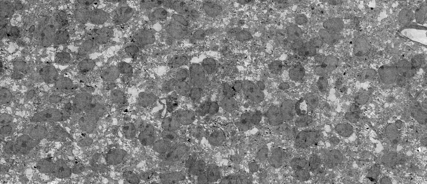
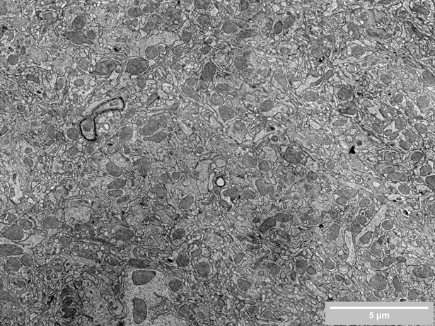
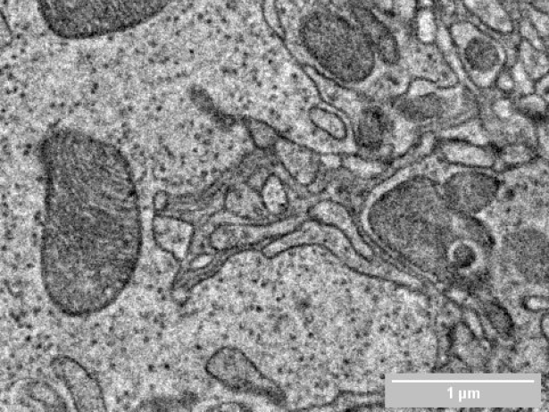
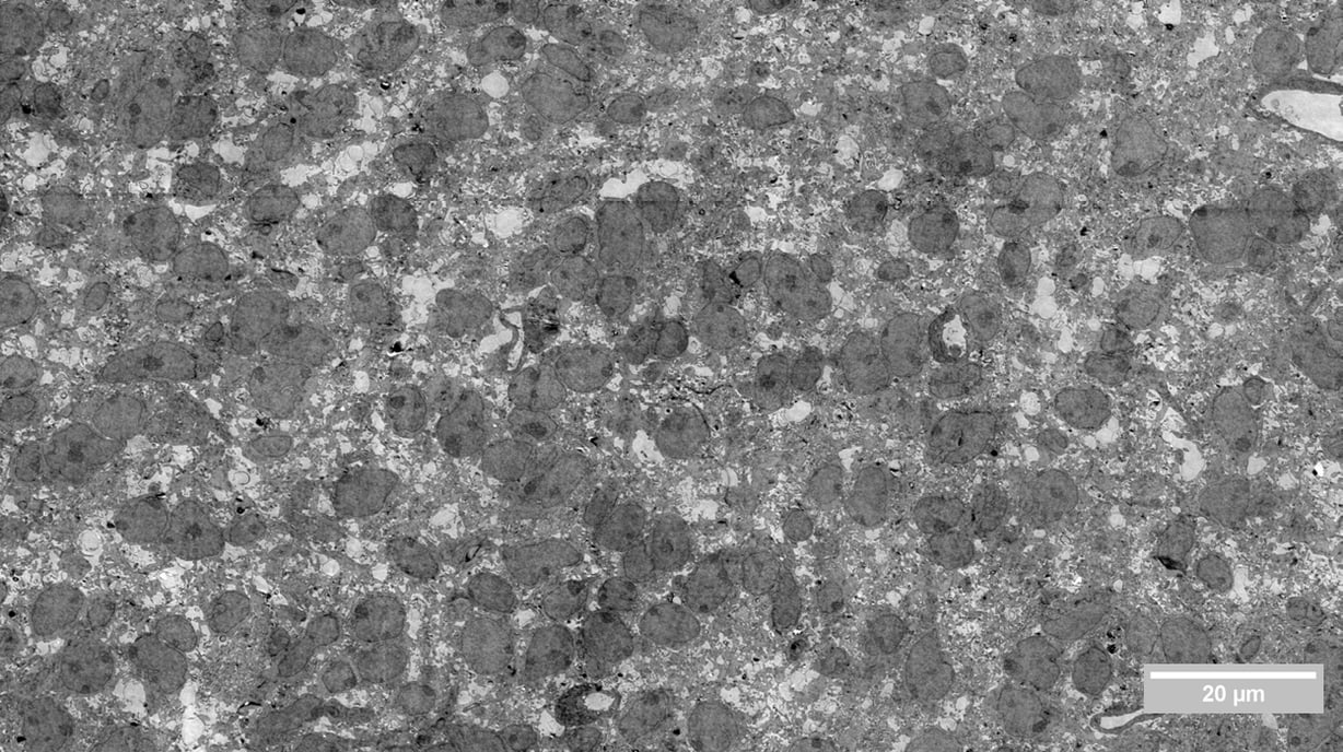
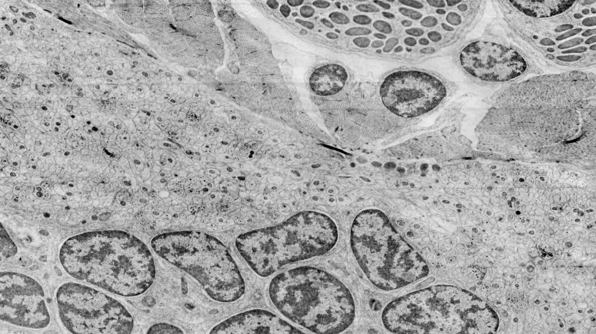
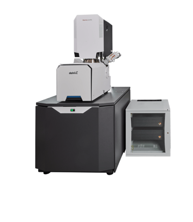
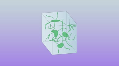
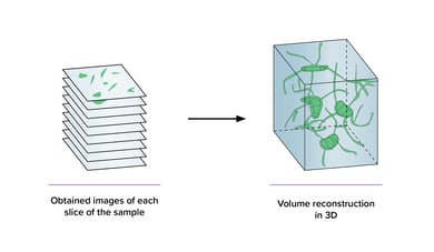
Electron imaging of all the sections is probably the most limiting factor in connectomics, as the throughput of an EM defines the size of the project. The time needed to record massive datasets in volume EM can go up to weeks, months or even years. To tackle this bottleneck we developed FAST-EM, an ultra-fast multibeam electron microscope, which accelerates the data collection steps required for connectomics. FAST-EM system combines high spatial resolution with high speed and automation and it speeds up existing projects and enables projects previously unfeasible with conventional scanning EMs.

What can you achieve with FAST-EM

FAST-EM significantly decreases the time needed to acquire large 3D images. This opens the way to larger projects, such as imaging entire brains for connectomics studies in reasonable amounts of time. At the same time, high-throughput imaging enables comparative studies, which were previously too time-consuming to be feasible.

  • Collect 3D EM datasets quickly
  • Acquire data with minimal operator supervision
  • Obtain your regions of interest pre-stitched and ready for your Z stack
Neuroscience_FI_Media Resize Neuroscience_FI_Media Resize_2
(Left) Drosophila brain – 60nm rOTO stained samples4 nm pixel size. (Right) Close-up view of complex cell-cell interfaces. Samples courtesy of Thomas Templier.

Use the right products to get the right results

Delmic Fast Imaging solutions consist of FAST-EM, an ultra-fast automated multibeam electron microscope. Learn today what it can do for your research.

Talk to an expert
Fast Imaging
Jacob Hoogenboom_IMG_9123-1

With FAST-EM, we can go to larger volumes. For instance, it could be used for a full model animal brain to map out all the neurons' connections

Dr. Jacob Hoogenboom

-

Faculty of Applied Sciences, TU Delft

Read customer story

Want to learn more?

Talk to one of our experts to learn how our solutions can support you in your research.

Get in touch
Talk-to-Expert_Marit-de-Beer_Avatar