Request pricing
Pathology

Visualize and analyze the anatomy of pancreatic cells

Study the pathophysiological mechanisms of diabetes type 1 with high throughput electron microscopy. Start capturing 3D data by imaging many subsequent sections of a sample with ease.

Learn about the solution
What we can help you to achieve
  • Resolve the intricate structures of organelles, cells, and tissues at nanometer-range resolution
  • Image significantly faster: acquire the necessary datasets in a matter of days
  • Visualize pathophysiological processes in 3D
  • Advance the research to find the cure

2021_FI_PancreasMegafieldIslet_MediaWithDescription

Speed up the understanding of Type 1 Diabetes

Type 1 diabetes occurs when insulin-producing beta cells present in the islets of Langerhans in the pancreas, are attacked and destroyed. Electron microscopy (EM) allows the identification of subcellular features of the beta cells, providing insight in the disease process. Traditionally, EM has had the drawback that acquiring images with a good signal-to-noise ratio is time-consuming, especially for large regions like entire islets of Langerhans. 

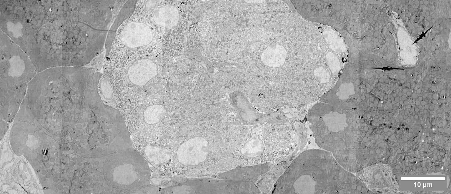
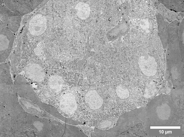
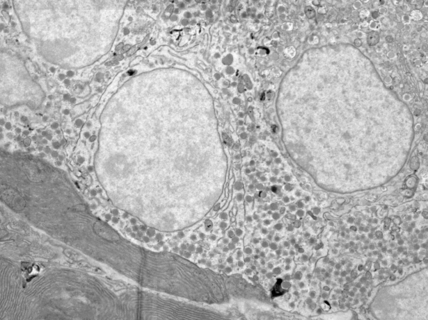
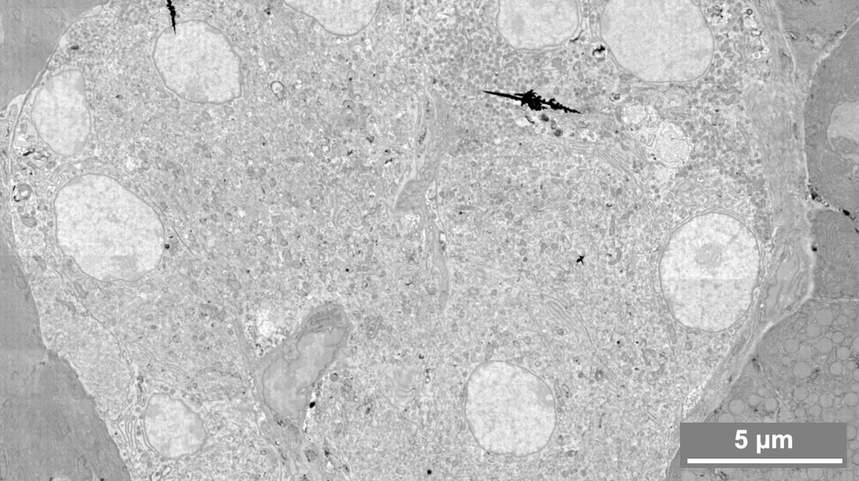
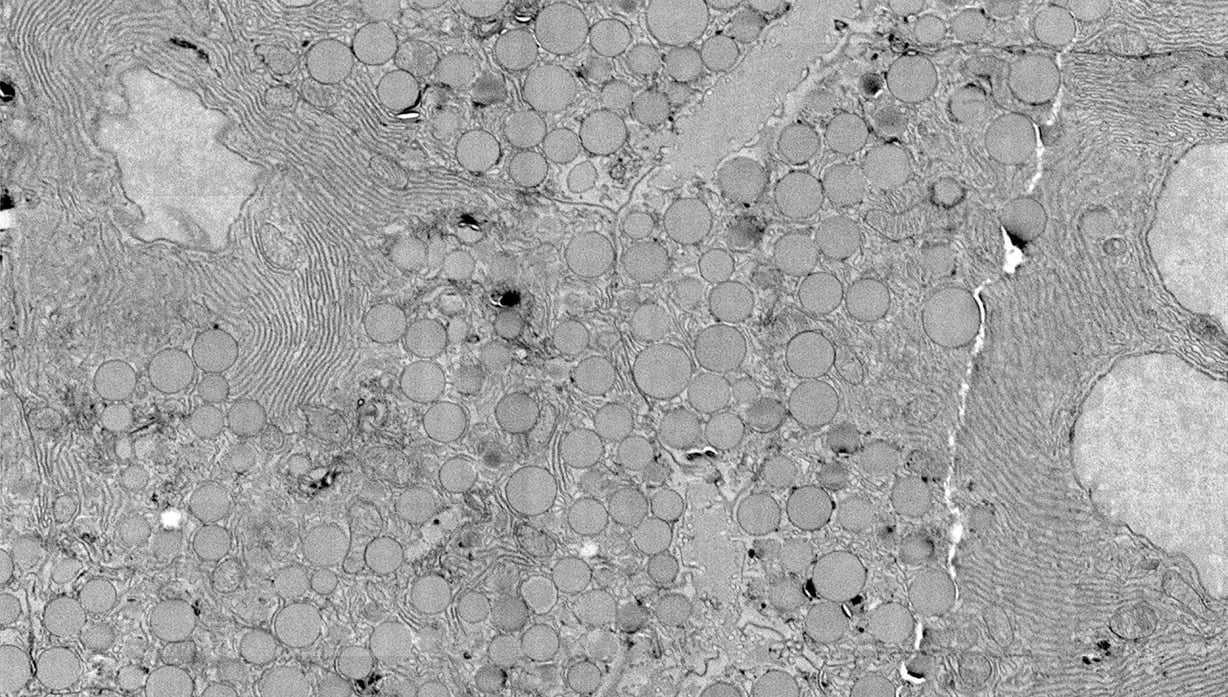
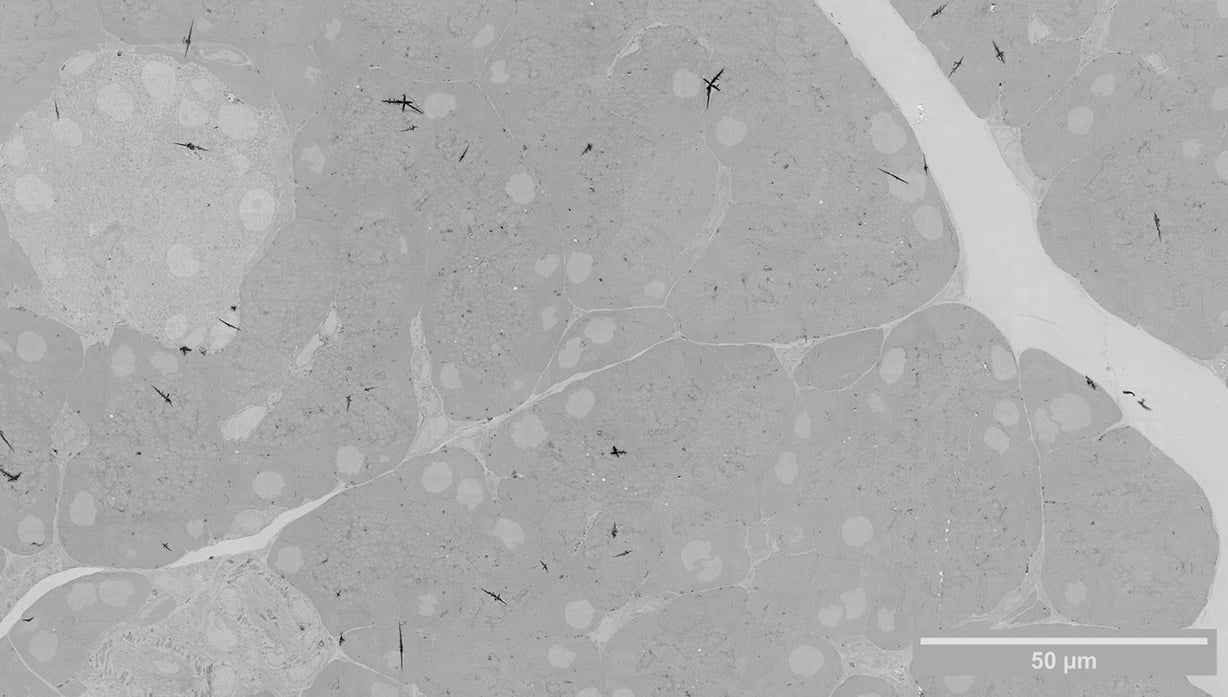
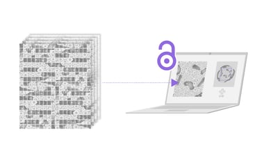
We created a powerful multibeam SEM, FAST-EM, which can rapidly visualize large regions, at nanoscale resolution. Its ability to image 100 times faster than a regular SEM and the high level of automation makes FAST-EM a perfect system for imaging the whole tissues or organs. Such high-throughput imaging enables comparative studies, where multiple samples need to be imaged and compared between healthy and diseased conditions. FAST-EM also enables a bias-free method of recording data: samples are imaged in their entirety, rather than specific parts relevant only to the research question.

What can you achieve with FAST-EM?

Traditional SEM techniques large-scale of 3D datasets are limited in their speed and often require significant experience to reach their full potential. A high level of automation and superior acquisition speed of FAST-EM, on the other hand, enables large-scale projects with minimal operator supervision. With FAST-EM you will be able to:

  • Acquire thousands of images of pancreatic tissue in a significantly shorter time
  • Analyze the microstructures of cells with high resolution
  • Perform comparative studies
  • Advance your research and get a clearer picture of the progression of diabetes
Diabetes_FI_Media Resize Diabetes_FI_Media Resize_2
Rat Pancreas. Sample courtesy: B.N.G. Giepmans and P. de Boer, UMCG. Field image of rat pancreas showing an islet of Langerhans (bottom left) and acinar cells (top right). Data captured using FAST-EM.
Diabetes_FI_Quote

We typically don’t take 3D images because if you have a large area, it takes us 6 hours for one 2D image. If you want to have a thousand sections, it will take a year. If you now have a very fast microscope, we can go for 3D imaging within a week of such a section.

Ben Giepmans, Principal Investigator

-

University Medical Center Groningen

Watch customer story

Use the right products to get the right results

Delmic Fast Imaging solutions consist of FAST-EM, an ultra-fast automated multibeam electron microscope. Learn today what it can do for your research?

Talk to an expert
Fast Imaging

Want to learn more?

Talk to one of our experts to learn how our solutions can support you in your research.

Get in touch
Style#1_Katherine_Avatar