Request pricing
Life sciences

Image and analyze biological samples with ease

Take your research to the next level with the power of automation and the unprecedented speed of the next generation electron microscopy solutions. Make statistically significant conclusions from your data easily, whether that is in pathology, neurobiology, connectomics, toxicology,  cell biology, or any other application in life sciences where both detail and context are important.

Learn about the solution
What can we help you with?
  • Image larger areas, more sections, bigger volumes, faster and with high resolution
  • Focus your time on analysis rather than on imaging your samples
  • Unlock the full potential of electron microscopy and obtain statistically significant answers to your research questions
Life Sciences_FI_Media with Description

Study complex and large biological samples

Electron microscopy (EM) is a powerful microscopy technique, which is widely used to examine the structures of cells, tissues, and organs, and to bring the researchers closer to answering fundamental questions. Thanks to the high resolution it offers, even small morphological differences can be identified and compared. Moreover, EM is increasingly being used in large-scale life science projects, either for volume imaging or large area mapping. In both cases, the desire to resolve nanoscale details is combined with the desire to place these observations into a larger context, which can cover a large area or span through a three-dimensional volume.

Such projects often face several limitations, particularly when multiple samples must be compared. Traditional EM workflows make acquiring statistically relevant data extremely time-consuming or even an unfeasible prospect. Fast electron microscopy brings much needed automation and high throughput to the table. It’s the ability to get both complete overview and in-depth data at nanometer resolution that makes fast electron microscopy so attractive. We developed a high-speed and fully automated electron microscope, FAST-EM. It eliminates the bottleneck created by the throughput of conventional electron microscopes.

Discover what you can achieve in your field

What we can offer:

Icon_FastEMViolet_Details and big picture
Get the details and the big picture

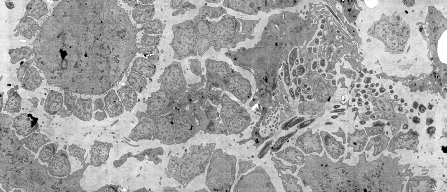
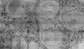
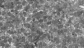
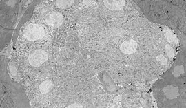
Collect nanoscale detail while retaining larger context of the sample

FeatureIcon_FI_SVG_270x270_Easy and fast
Boost productivity and focus on data analysis

Achieve high, sustained, throughput and image hundreds of sections with high levels of autonomy

FeatureIcon_FI_SVG_270x270_Measure faster
Easily achieve statistical impact with your EM data

Faster imaging enables collection of data from more samples, conditions, or time points

FeatureIcon_FI_SVG_270x270_Customer support
Support from our application experts

Get the most out of your instrument with our guidance and help

Jacob Hoogenboom_IMG_9123-1

Now we can go to more samples, to larger volumes, and get to inter-sample variability, which will open many new questions on the technology side and the biology side

Dr. Jacob Hoogenboom

-

Faculty of Applied Sciences, TU Delft

Read case study

Want to learn more?

Talk to one of our experts to learn how our solutions can support you in your research.

Get in touch
Style#1_Katherine_Avatar