Request pricing
Cancer research

Study cancer processes

Integrated CLEM enables the study of rare and transient events occurring within the diseased cell at high resolution. This technique has great potential for shedding light on the process by which cancer develops and spreads.

Download white paper
What we can help you achieve:
  • Identify a region of interest rapidly
  • Collect highly detailed data in vivo easily
  • Understand how the tumor cell operates within the context of surrounding tissue
BSE image of an individual cell expressing GFP-C1

The effects of cancer at the nanoscale

Cancer research is an important field which investigates the causes of cancer and develops methods for treatment. The development and spread of cancer is a dynamic process which starts within the genes of a cell or a group of cells. Understanding these processes, which happen on a nanoscale level, is essential to developing methods that can limit the spread of cancer to other parts of the body.

Correlative light and electron microscopy (CLEM) has consistently  been gaining popularity in the field of cancer research. The Delmic integrated CLEM  enables the observation of very specific biochemical changes at the nanoscale by combining the labelling power offered by fluorescence microscopy with the high-resolution contextual information from the scanning electron microscope.

What can you achieve with integrated CLEM?

While using the SECOM, scientists were able to examine the loss of the SMAD4 gene in transfected and cultured colorectal cancer cells. The changes in cell shape and small cell protrusions were clearly observed, for instance, invadopodia degrades the extracellular matrix and contributes to metastasis. Researchers argue that it was only with a system that could perform high-aperture fluorescence and electron microscopy simultaneously that these changes in the cells could be observed.

Superresolution image showing localisation of YFP-A3 in a HeLa cell Wide field image showing localisation of YFP-A3 in a HeLa cell
Superresolution image of a cell reconstructed from a sequence of approximately 30,000 images, demonstrating the improvement in the precision of YFP-A3 localisation with respect to wide field images
Dr. Lucy M. Collinson, Francis Crick Institute, London, United Kingdom

At our EM Science Technology Platform, [...] we use the SECOM SR platform to perform high accuracy CLEM experiments on resin-embedded cells and tissues containing fluorescent proteins. Implementing superresolution localisation microscopy in the SECOM SR using the blinking of standard fluorescent proteins in-resin takes us to the next level...

Dr. Lucy M. Collinson and Dr. Christopher J. Peddie

-

Francis Crick Institute, London, United Kingdom

Read case study

Use the right products to get the right results

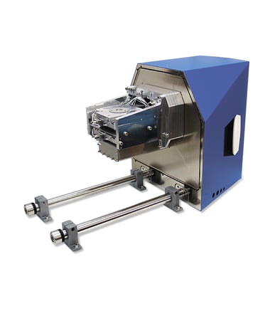
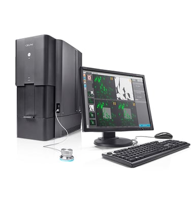
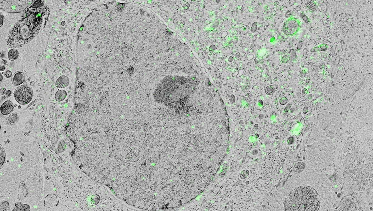
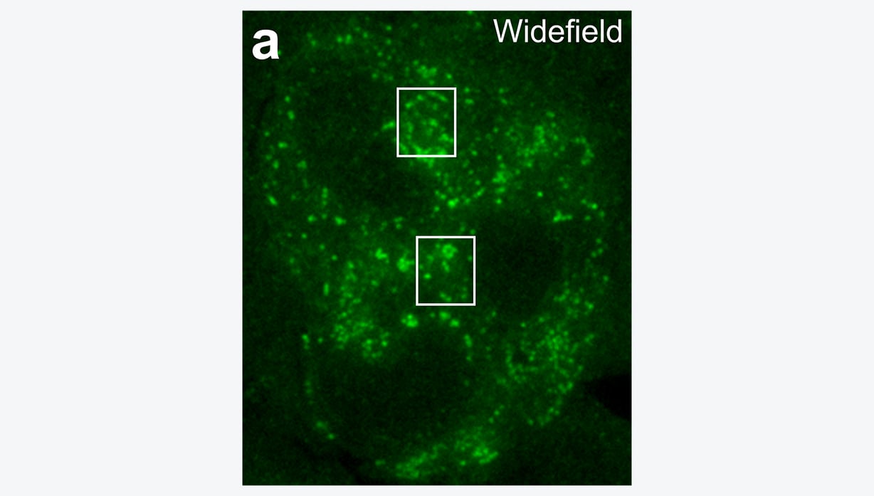
Delmic CLEM solutions consist of powerful integrated correlative light and electron microscopy systems, which can help you understand more about your biological sample.

Talk to an expert
Solutions empowering cancer research

Talk to an expert

Get in touch with our Sales representative Kevin Homberg to get more information.
Call +31 (0)15 744 01 58 or book a meeting in his agenda.

Book a meeting
CTA Bar - Kevin - PNG