Request pricing
Cell biology

Study cells to understand underlying mechanisms of life

Combining light and electron microscopy allows studying the ultrastructure of cells with high-resolution electron microscopy, while simultaneously performing fluorescence imaging to reveal valuable information about cellular function from different parts of the cell. The next step of studying cells is cryo-ET, which can shed light on the connections between different intracellular membrane systems by membrane contact sites. 

Download application note
What we can help you to achieve
  • Identify the region of interest rapidly

  • Understand how the cell operates within the context of surrounding tissue

  • Collect highly detailed data in vivo easily and save time with automated image overlay
  • Improve lamella-preparation and overall cryo workflow with integrated cryo solutions
header Localization of the lipid diacylglycerol within cellular membranes of Hela cell expressing GFP-C1

Fluorescence and electron images of cells without sample transfer

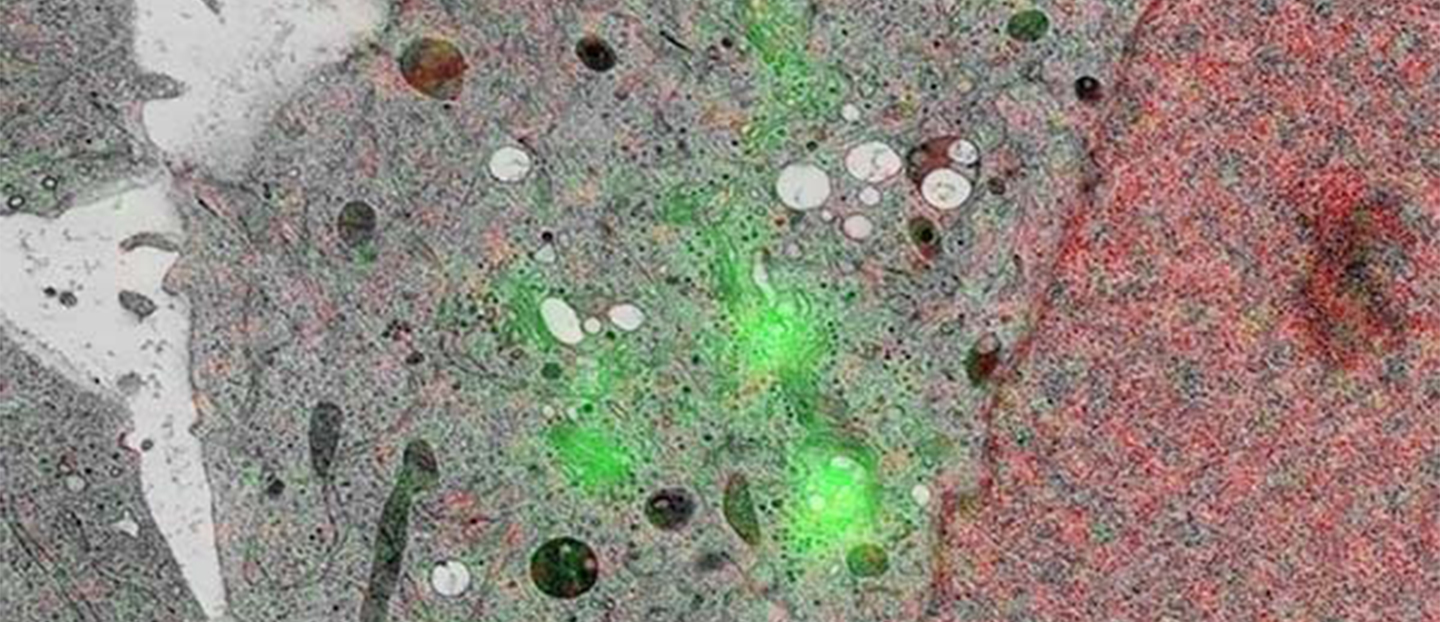
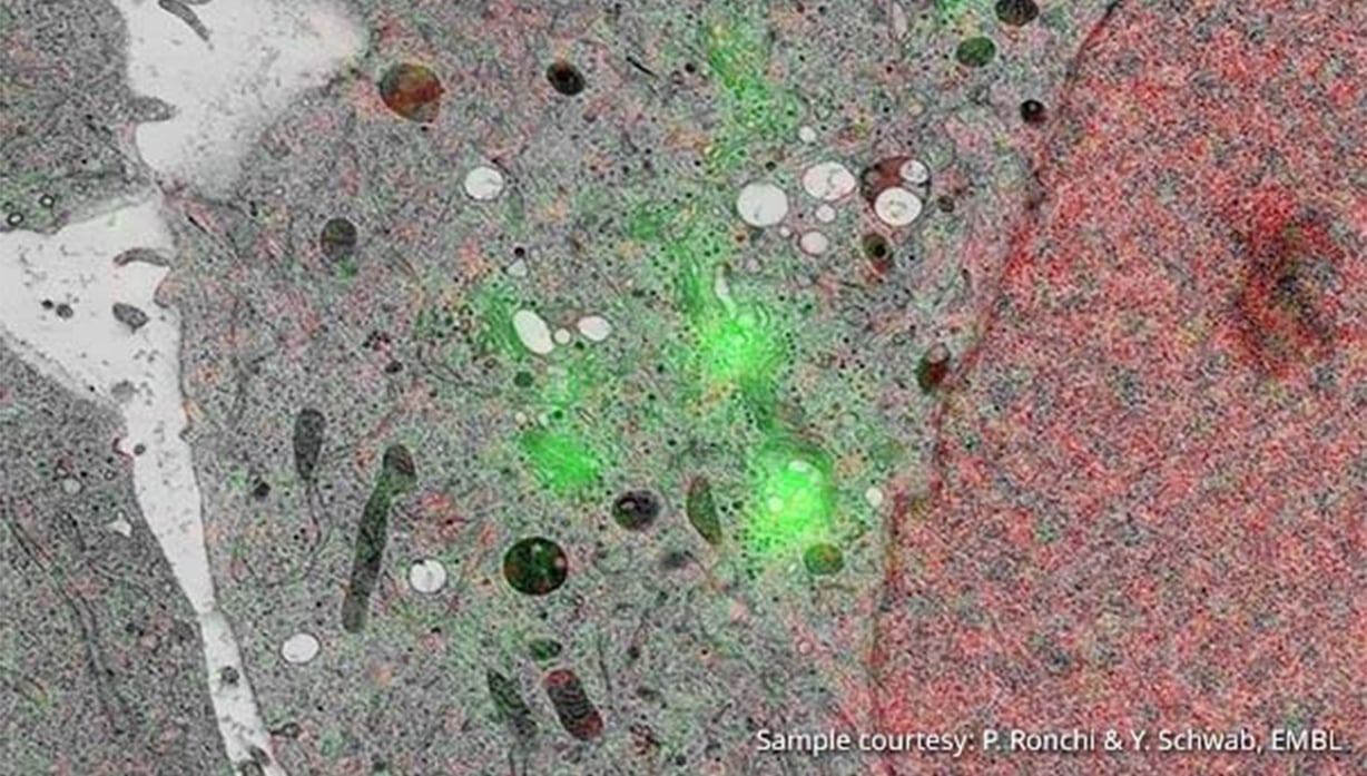
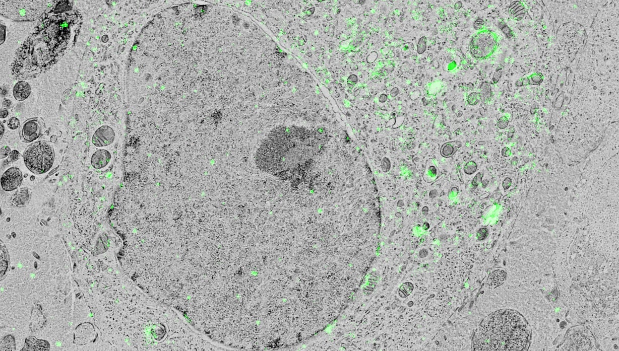
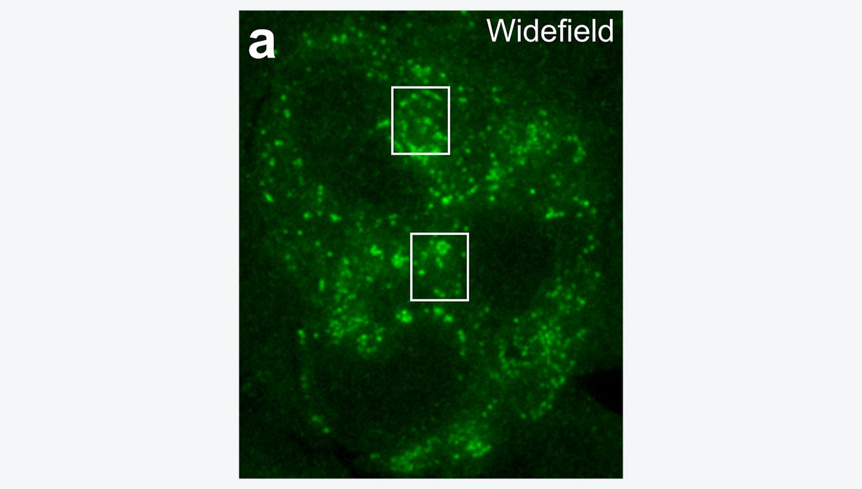
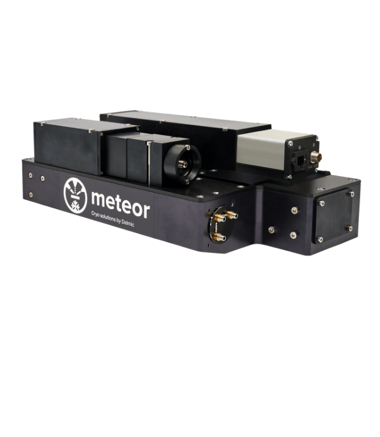
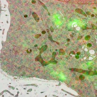
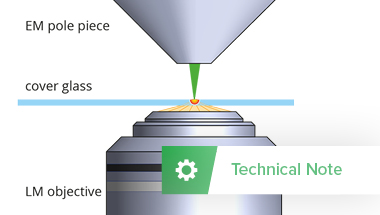
Integrated CLEM combines the two imaging methods EM and FM, to offer a powerful streamlined and automated acquisition of high resolution CLEM images in the SEM. The platform is a retro fit compatible with SEM’s from the entire range of manufacturers. In iCLEM, the cells of interest can be identified using FM. Subsequently, a high magnification EM image can be acquired at the desired location to reveal structural details at high resolution. Following this, the two acquired images will be overlayed using the high accuracy automated overlay procedure. It therefore offers seamless CLEM imaging with no sample transfer or manual overlay, providing high quality data in a user-friendly manner, ensuring that you can focus on analyzing data rather than on acquiring it.

Integrating a fluorescence light microscope into the FIB/SEM can optimize the cryo-ET workflow. Using an integrated FLM in the FIB/SEM means that ROI identification by FLM, SEM imaging, FIB milling and ROI confirmation by FLM can all be done in the same system without the need to handle the sample, thus increasing the success rate of the cryo-ET workflow considerably. No need for dangerous and damaging transfer steps.

What can you achieve with integrated CLEM?

The integrated CLEM solution allows you to image the cell(s) of interest with both EM and FM in one integrated system, streamlining the workflow and avoiding sample damage and contamination. HeLa cells transfected with GFP-C1 have been imaged to achieve super-resolution molecular localisations of ~ 85 nm, enabling the high-precision correlation of fluorophore-labelled macromolecules to cellular ultrastructure.

Superresolution image of a cell demonstrating the improvement in the precision of YFP-A3 localisation Wide field image of a cell Wide field image showing localisation of YFP-A3 in a HeLa cell
Superresolution image of a cell reconstructed from a sequence of approximately 30,000 images, demonstrating the improvement in the precision of YFP-A3 localisation with respect to wide field images

Use the right products to get the right results

Delmic CLEM solutions consist of powerful integrated correlative light and electron microscopy systems, which can help you understand more about your biological sample.

Talk to an expert

Talk to an expert

Get in touch with our applications specialist Caspar Jonker to get more information.
Call +31 (0)15 744 01 58 or book a meeting on his agenda.

Book a meeting
Caspar application pages