Request pricing
Neuroscience

Trace neurons throughout the brain

Understanding the wiring of the brain requires visualizing its constituent cells with electron microscopy, the only technique that can visualize the nanoscale details that define individual cells. By using high-throughput electron microscopy, these details can be collected on a large scale, providing data for a full map of all interactions in the brain, helping researchers examine processes like behaviour, memory, and decision making.

Read application note
What we can help you to achieve
  • Image large volumes of brain tissue
  • Relate morphology of neurons with their function
Electron micrographs of songbird brain in neuroscience

Overcoming the data challenges of connectomics

One of the most fascinating challenges in neuroscience is the mapping of the complete connectivity of the brain. Recent years have seen a rapid expansion in the field of connectomics, the study of tracing and understanding this connectivity map. One of the methods of choice in the field of connectomics is 3D electron microscopy (3D EM). 3D EM enables imaging of large volumes of brain tissue at nanoscale resolution, to study the structures that define individual brain cells and interactions with other cells, even at long distances.

Delmic’s Fast EM drastically accelerates the data collection steps required for connectomics, in a system that combines that combines high spatial resolution with high speed and automation. Fast EM speeds up existing projects and enables projects previously unfeasible with conventional scanning EMs.

What can you achieve with Fast EM Imaging?

Delmic’s Fast EM solution is highly suited for neuroscience thanks to its high sustained throughput. The high level of automation enables large-scale 3D EM workflows with minimal operator supervision. Volumes of interest are rapidly collected, which enables neuronal tracing over large distances and a better understanding of cellular interactions in the brain.

  • Collect 3D EM datasets quickly
  • Acquire data with minimal operator supervision
  • Larger volumes of data within the same time
Electron micrographs of songbird brain in neuroscience Electron micrographs of songbird brain in neuroscience
Electron micrographs of songbird brain. Electron microscopy enables the reconstruction of synapses by resolving synaptic vesicles and post-synaptic densities at high resolution, making it one of the methods of choice for connectomics. Sample courtesy of Thomas Templier (ETH Zurich, Switzerland).
illustration of Fast electron microscopy workflow

Workflow for using 3D EM for connectomics.

Shown are the preparatory steps of before imaging, the imaging cycle used to acquire image data from all tiles in the dataset, and the reconstruction of image data to form a volume of the brain.

Publication

Pieter Kruit, Wilco Zuidema. "A Dedicated Multi-Beam SEM for Transmission Imaging of Thin Samples." Microscopy and Microanalysis 25.S2 (2019): 1034-1035.

Choose the right product for your research

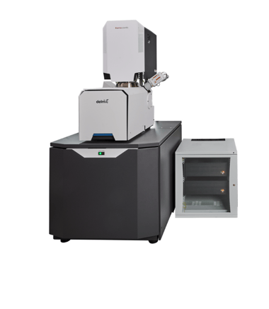
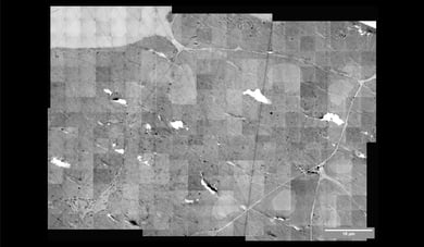
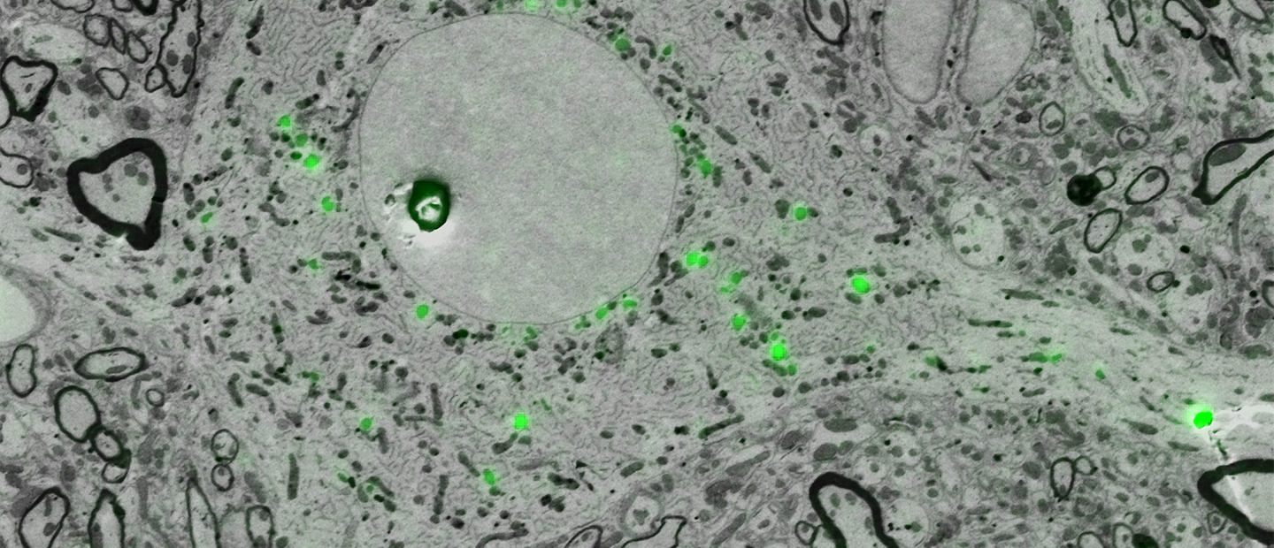
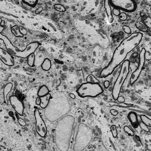
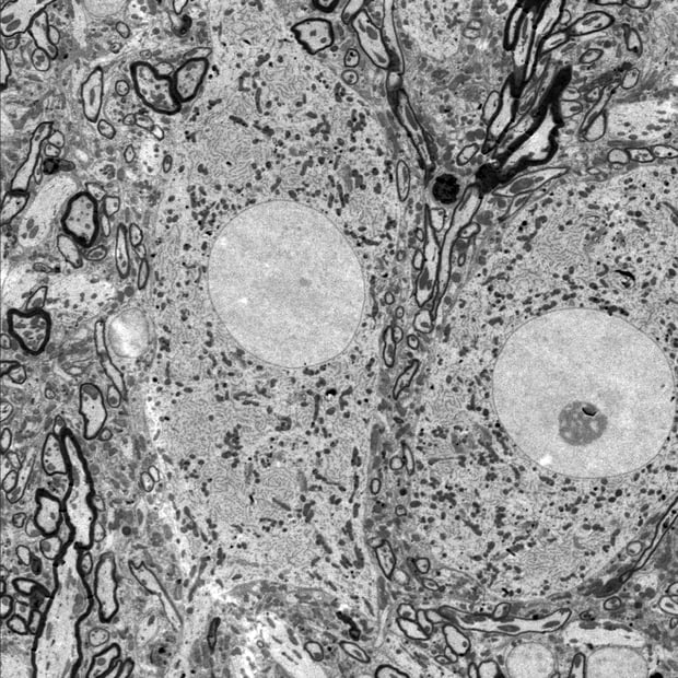
Delmic CLEM solutions consist of powerful integrated correlative light and electron microscopy systems, which can help you understand more about your biological sample.
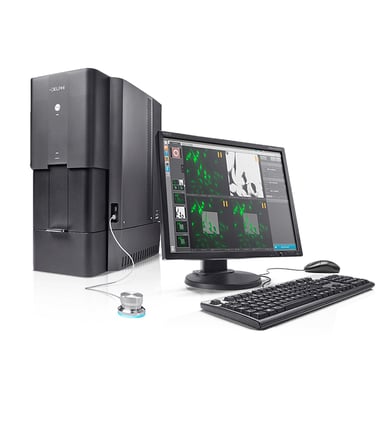
Go to Fast EM technique page
Products empowering neuroscience

Talk to an expert

Get in touch with our  sales representative Kevin Homberg to get more information.
Call +31 (0)15 744 01 58 or book a meeting on his agenda.

Book a meeting
CTA Bar - Kevin - PNG