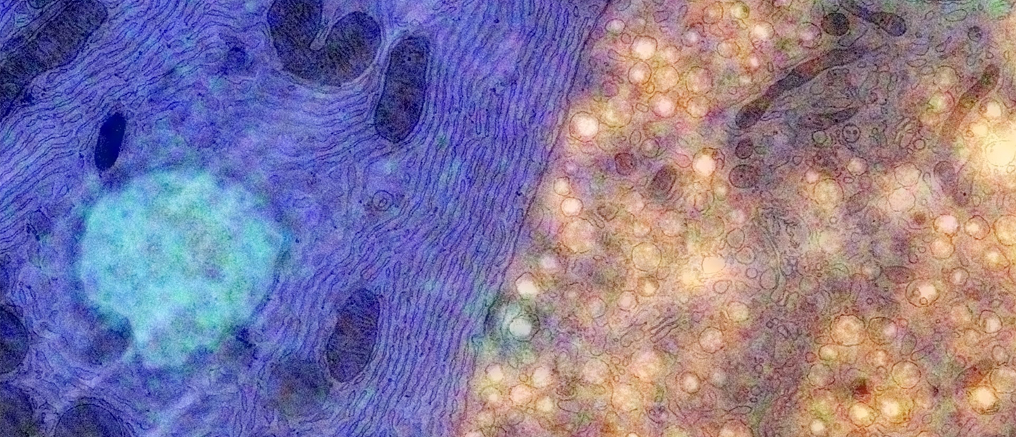
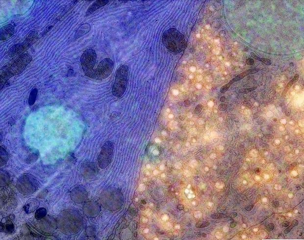
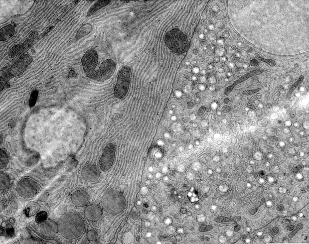
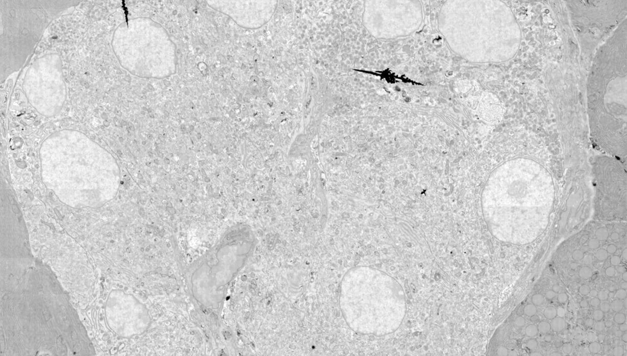
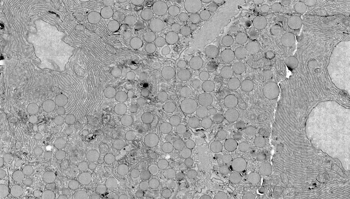
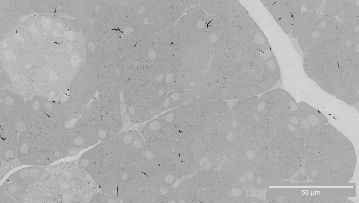
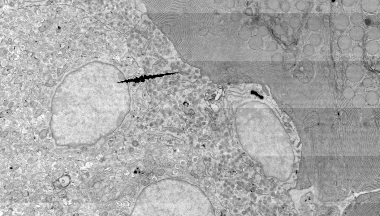
Type 1 diabetes occurs when insulin-producing beta cells present in the islets of Langerhans in the pancreas, are attacked and destroyed. Electron microscopes (EM) allow the identification of subcellular features of the beta cells, providing insight in the disease process. Traditionally, EM has had the drawback that acquiring images with a good signal-to-noise ratio is time-consuming, especially for large regions like entire islets of Langerhans.
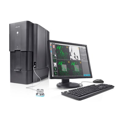
Delmic has developed two solutions that accelerate the use of EM in diabetes research. The first, integrated Correlative Light and Electron Microscopy (CLEM), combines FM and EM in one instrument, enabling the user to perform CLEM in a streamlined and automated manner. Integrated CLEM first rapidly images cells over a large field of view using FM, to identify the area of interest