Request pricing
Diabetes

Image fluorescently labelled beta cells

Delmic offers several solutions that enable you to accelerate research into Type 1 diabetes. Correlative light and electron microscopy is perfect to combine functional and structural data, while Fast EM provides a high-throughput pipeline to image entire sections or compare samples.

Download application note
What we can help you to achieve
  • Simultaneously acquire high resolution correlative image data of the islets of Langerhans
  • Rapidly acquire large-scale data from entire islets of Langerhans
  • Examine insulin-producing cells with extremely simplified workflows
  • Visualize pathophysiological processes in 3D with automated sample preparation and high throughput
20151208-162429_fig4_FMEM_RES

Speed up the understanding of Type 1 Diabetes

Type 1 diabetes occurs when insulin-producing beta cells present in the islets of Langerhans in the pancreas, are attacked and destroyed. Electron microscopes (EM) allow the identification of subcellular features of the beta cells, providing insight in the disease process. Traditionally, EM has had the drawback that acquiring images with a good signal-to-noise ratio is time-consuming, especially for large regions like entire islets of Langerhans. 

Delmic has developed two solutions that accelerate the use of EM in diabetes research. The first, integrated Correlative Light and Electron Microscopy (CLEM), combines FM and EM in one instrument, enabling the user to perform CLEM in a streamlined and automated manner. Integrated CLEM first rapidly images cells over a large field of view using FM, to identify the area of interest 

based on cellular function, followed by selective high-resolution EM imaging at that location. In this way, both functional and structural data is acquired in a seamless manner.

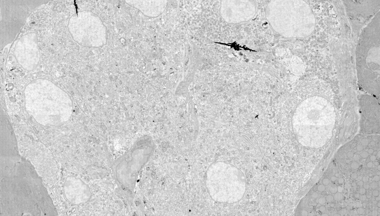
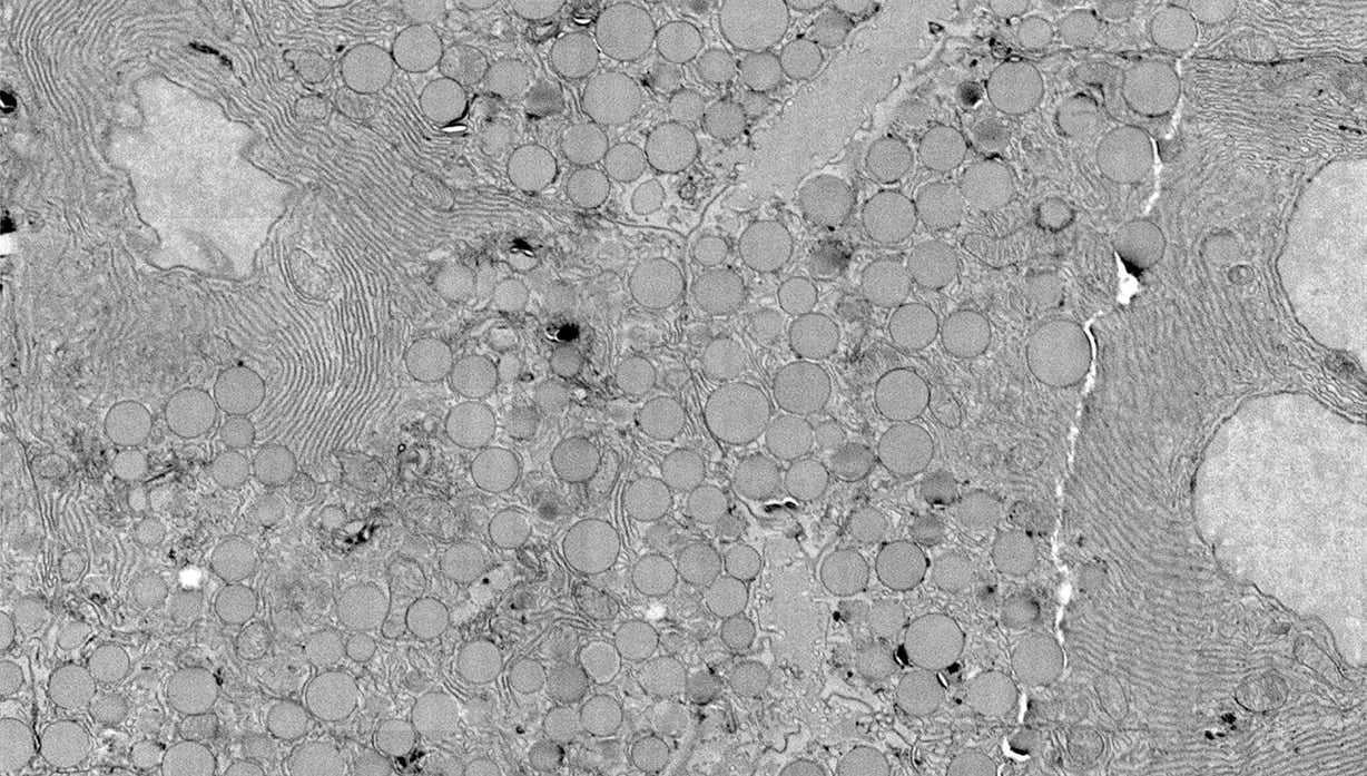
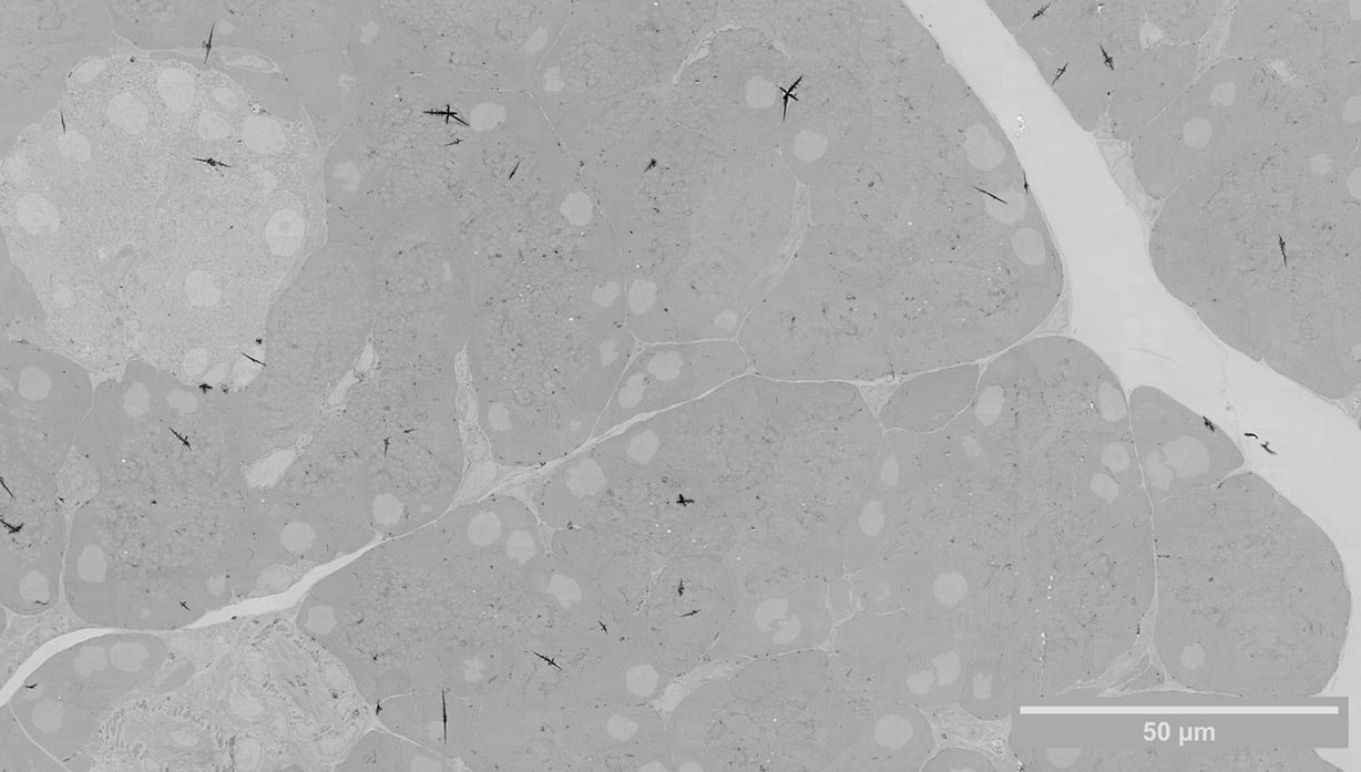
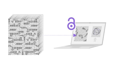
A second solution, Fast EM, harnesses the increased throughput of a multibeam scanning EM to rapidly visualize large regions, at nanoscale resolution. High-throughput imaging enables comparative studies, where multiple samples need to be imaged and compared between healthy and diseased conditions. Fast EM also enables a bias-free method of recording data: samples are imaged in their entirety, rather than specific parts relevant only to the research question. In addition, the high throughput of FAST-EM enables rapid 3D visualization of pancreatic tissue, with which the pathophysiological processes can be examined in three dimensions.

What can you achieve with Delmic's solutions?

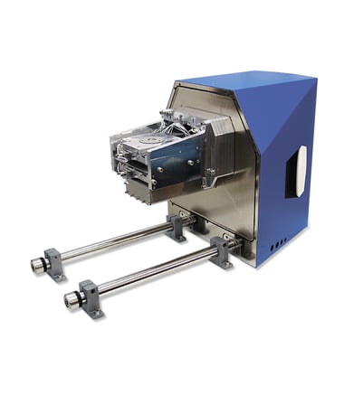
Integrated CLEM combines the two imaging methods EM and FM, to offer a powerful streamlined and automated acquisition of high resolution CLEM images in the SEM. The platform is a retrofit compatible with SEM’s from the entire range of manufacturers.

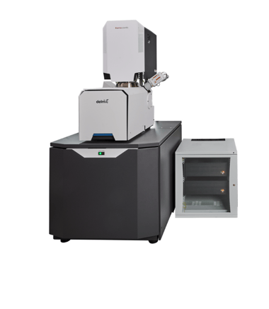
Fast EM is a system dedicated to high throughput EM. The system’s speed and automation provide a stable workflow that requires minimal operator interaction.

  • Understand the changes taking place in the beta cells due to diabetes
  • Simultaneously identify the morphology and cellular function
  • Provide data for research into the fundamental cause of type 1 diabetes
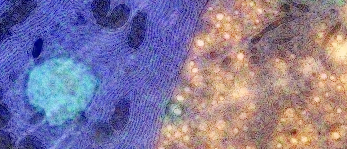
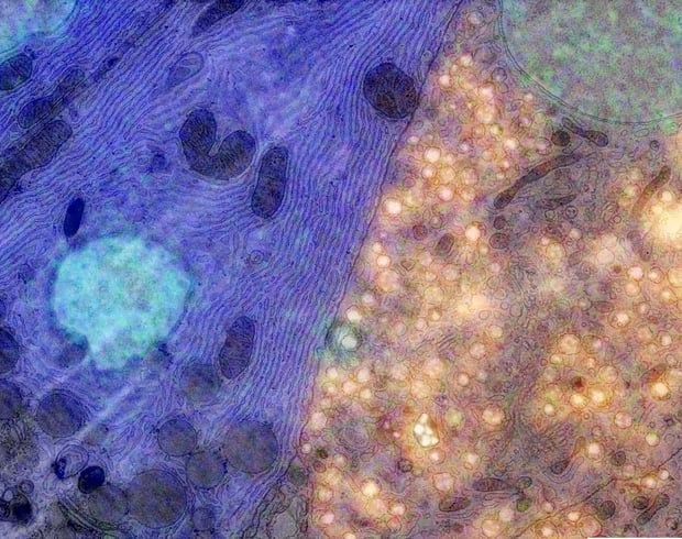
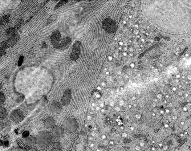
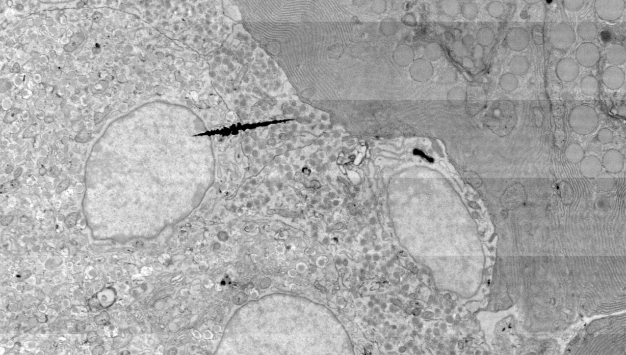
simultaneously acquired correlative image of the islets of Langerhans of Rat Pancreas EM image of the islets of Langerhans of Rat Pancreas
Rat Pancreas. Data captured using the EM and SECOM system. Sample courtesy: B.N.G. Giepmans & P. de Boer, UMCG. Simultaneously acquired correlative image of the islets of Langerhans imaged on the Delmic SECOM integrated with an FEI Verios 460 Scanning Electron Microscope. The insulin (beta cells) are labelled in orange with Alexa Fluor 594, with the ultrastructure is visible in the EM contrast. The guanine quadruplexes are labelled in green with Alexa Fluor 488, and the nucleus is labelled in blue with Hoechst.

Use the right products to get the right results

Delmic CLEM solutions consist of powerful and user-friendly CLEM systems, which can help you learn fast and more about your biological sample. Choose the product best suited to your research questions!

Talk to an expert
CLEM and Fast Imaging solutions

Talk to an expert

Get in touch with our sales representative Kevin Homberg to get more information.
Call +31 (0)15 744 01 58 or book a meeting on his agenda.

Book a meeting
CTA Bar - Kevin - PNG